The Brain’s Repair Crew May Not Be Neurons
When people think about brain repair, neurons usually get the spotlight, but they rely on support cells called glia to maintain the brain’s environment. In July 2026, researchers reported an unexpected form of astrocyte regeneration in the adult mouse brain after focal astrocyte loss. Surviving astrocytes near the damaged region reorganized, proliferated and produced daughter-cell nuclei that travelled through extended cellular processes into the depleted area, helping rebuild the local astrocyte network. The finding suggests that the adult brain may have more capacity for certain forms of glial repair than previously appreciated and reveals another layer of cellular plasticity. However, it remains an experimental finding in mice and does not mean that damaged human brain tissue can fully regenerate after major injury.
Meet the Astrocytes: The Brain’s Multitasking Support Cells
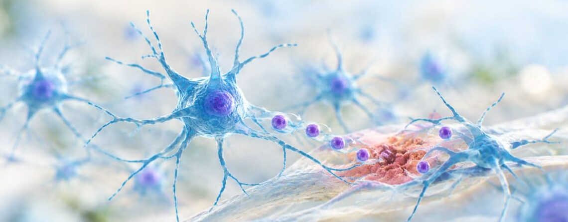
Astrocytes are star-shaped glial cells distributed throughout the central nervous system. Their responsibilities include maintaining the chemical environment around neurons, supporting energy metabolism, helping regulate blood flow, participating in neurotransmitter recycling and contributing to the blood-brain barrier and tissue homeostasis.
They are also dynamic cells. Following injury, astrocytes can undergo reactive astrogliosis, changing their shape, molecular profile and interactions with neighbouring cells. Historically, this response was often viewed primarily through the idea of a “glial scar”; a barrier that could restrict regeneration. Modern research has made that picture considerably more nuanced: reactive astrocytes can also contain damage, protect surviving tissue and participate in repair-related processes.
The 2026 discovery adds something different: under a particular pattern of localized astrocyte loss, surviving astrocytes can participate in actual repopulation of the depleted territory.
Overturning the Old Assumption About Glial Regeneration
The new study examined what happens when astrocytes disappear from a small, defined region of the adult mouse cortex. Researchers used an antibody directed against aquaporin-4 (AQP4) to selectively create focal astrocyte loss. AQP4 is a water channel strongly associated with astrocytes and is also the target of pathogenic antibodies in neuromyelitis optica spectrum disorder, or NMOSD.
What followed was striking. Astrocytes surrounding the lesion did not simply remain at its edge. They changed their structure, extended processes toward the empty territory, underwent proliferation and developed a temporary multinucleated state. Over subsequent weeks, nuclei from newly generated daughter cells progressively occupied areas that had lost their astrocytes.
This does not overturn the broader reality that the adult brain has limited regenerative capacity compared with tissues such as skin or liver. Rather, it overturns a narrower assumption: that mature astrocyte networks are incapable of rebuilding themselves after spatially restricted cell loss.
A Repair Strategy That Looks Different From Ordinary Cell Division
Ordinary cell division is usually described as a cell growing, duplicating its genetic material and separating into daughter cells. The new observations reveal a more unusual spatial arrangement.
In the mouse model, astrocytes around the damaged area proliferated but did not immediately place every daughter nucleus beside its original cell body. Instead, newly formed nuclei became associated with elongated cellular processes extending toward the vacant region. The nuclei then gradually shifted into territories that had become empty.
In other words, the cellular infrastructure appears to reach into the damaged space first, while nuclei subsequently occupy that territory. The study describes a prolonged multinucleated intermediate state before nuclei become positioned within the previously depleted area. That is fundamentally different from imagining a new astrocyte simply being born exactly where another astrocyte was lost.
The Mechanism of Long-Distance Nuclear Movement
The word “migration” can be misleading if it creates the impression that complete astrocytes detach and crawl independently into the lesion. That is not what the researchers observed. Instead, the nuclei themselves were displaced through the existing or newly remodelled cellular architecture. Astrocytes extended polarized processes into the depleted region, creating elongated routes through which the newly generated nuclei could move. The cellular body and broader astrocytic processes remained part of the surrounding network rather than an entire cell travelling independently across the lesion.
This distinction is one of the most fascinating aspects of the finding. The repair process appears to exploit the highly branched architecture of astrocytes themselves, turning their normally extensive cellular geometry into a route for rebuilding the local network.
Only the Nuclei Move: Not Whole Astrocytes
One of the most important details to get right is that researchers did not observe whole astrocytes marching into the lesion. The striking movement involved newly generated daughter-cell nuclei. Those nuclei shifted through long cellular extensions toward previously unoccupied territories. Eventually, the depleted region became repopulated and the characteristic astrocyte “tiling” of neighbouring territories was re-established. This is why the discovery is better described as nuclear translocation during astrocyte repopulation rather than conventional cell migration.
How Did Scientists Watch the Process in Real Time?
The researchers combined two powerful approaches: longitudinal in vivo two-photon microscopy and molecular mapping of gene activity.
Two-photon microscopy allowed the team to repeatedly observe labelled cells inside the brains of living mice over a period of several weeks. Instead of examining one animal at one time point and reconstructing what might have happened, researchers could follow the same repair process as it unfolded. The technique provided a window into changing cell shapes, processes and nuclear positions in living tissue.
They paired this imaging with spatiotemporal transcriptional profiling, including spatial transcriptomics, to identify molecular changes associated with the repair process. This allowed the researchers to connect what the cells were physically doing with changes in gene expression. That combination is important. Microscopy showed the movement and structural remodelling; molecular profiling helped reveal the biological state accompanying the repair.
Rebuilding a Functional Glial Network
The goal of the process was not simply to fill an empty patch. Astrocytes normally occupy organized territories and maintain extensive interactions with neurons, blood vessels and neighbouring glial cells. As the damaged region was repopulated, the researchers observed restoration of astrocyte organization. The injury-associated transcriptional response also gradually resolved as the astrocyte network was re-established.
This suggests that regeneration involves more than producing replacement cells. The brain must also restore the architecture and relationships that allow those cells to function as a network. That distinction matters for regenerative medicine. Replacing a cell is not necessarily equivalent to restoring tissue function. A successful repair process needs cells to occupy the right places and reconnect with the surrounding biological environment.
What Conditions Can Cause Astrocyte Loss?
Astrocyte loss or dysfunction can occur in several neurological settings. Traumatic brain injury, ischemic injury, neuroinflammatory diseases, infections and autoimmune disorders can all disrupt astrocytes, although the pattern and extent of damage differ substantially between conditions.
NMOSD is particularly relevant to this study because many patients have antibodies against AQP4. These antibodies can damage astrocytes and contribute to inflammatory lesions in the central nervous system. Experimental work has demonstrated rapid astrocyte depletion following AQP4-antibody exposure, with subsequent injury to other neural structures.
The new research used an AQP4-antibody-mediated mouse model specifically because it creates a controlled example of focal astrocyte loss relevant to NMOSD pathology. It should not be interpreted as showing that people with NMOSD naturally undergo the exact same repair sequence observed in mice.
Why This Matters for Neuroregeneration
Neuroregeneration is often discussed as though the central challenge is simply replacing dead neurons. The astrocyte findings broaden that picture. Neurons depend on their surrounding cellular environment. If astrocytes are lost, the resulting disruption can affect metabolic support, neurotransmitter regulation, water balance, vascular interactions and tissue stability. Restoring astrocyte organization could therefore become an important component of repairing neural tissue even when lost neurons themselves cannot be replaced.
The 2026 study does not demonstrate functional recovery from a neurological disease in humans. Its importance is that it identifies a previously underappreciated cellular repair mechanism that could be investigated as one component of future regenerative strategies.
Molecular Targets for Future Therapeutics
The researchers identified temporary changes in gene expression associated with the repair response. These molecular signatures provide potential starting points for investigating how astrocytes switch from a damaged or reactive state into a regenerative one.
However, a molecular pathway appearing during regeneration does not automatically become a safe drug target. Future work must determine which signals initiate nuclear movement, which regulate process extension, how proliferation is controlled and how the repair response eventually stops.
The researchers also found that the observed remodelling was not dependent on connexin 30/connexin 43 signalling or the vascular niche, providing useful constraints on the mechanism. The extent of astrocyte loss itself appeared to influence whether this unusual remodelling response occurred.
Targeted Clinical Applications: Where Could This Lead?
The most immediate conceptual applications include disorders involving focal astrocyte loss, particularly NMOSD, and potentially selected forms of traumatic brain injury. But “potential application” is very different from an available treatment.
For NMOSD, preventing the autoimmune attack remains clinically central. A future regenerative therapy might theoretically complement disease control by helping damaged tissue rebuild its astrocyte environment after an attack. For traumatic brain injury, the situation is more complicated because injury affects neurons, axons, blood vessels, oligodendrocytes, immune cells and extracellular structures in addition to astrocytes. The repair mechanism may therefore prove useful as one piece of a larger regenerative strategy rather than a universal solution for brain injury.
An Important Reality Check: Mouse Repair Is Not Human Brain Regeneration
The discovery is exciting precisely because it reveals something unexpected in the adult mouse brain. But the evidence currently stops there for the core mechanism.
The researchers did observe astrocytic features resembling the repair response in tissue from patients with NMOSD, which provides an important human connection. Nevertheless, the detailed nuclear-translocation process was experimentally demonstrated in mice.
There is also a major difference between repairing a carefully controlled focal lesion and recovering from a large human stroke, severe traumatic brain injury or widespread neurodegenerative disease. Human injuries may involve much larger areas, complex inflammation, vascular damage and irreversible neuronal loss.
So the discovery should be described as evidence of previously unrecognized regenerative capacity in adult mammalian astrocyte networks, not proof that the human brain can simply regenerate itself.
Recent Research Highlights
The July 2026 Nature Neuroscience study is the key breakthrough: mature perilesional astrocytes responded to focal loss by proliferating, extending processes and repositioning daughter-cell nuclei into depleted territories. The response restored astrocyte tiling within weeks in the mouse model and was accompanied by a temporary injury-related transcriptional program.
The broader 2026 astrocyte literature is also moving away from viewing glial cells as passive support. A 2026 review emphasizes that astrocytes display highly diverse reactive states depending on disease, injury stage and context, while recent experimental work has shown that astrocytes can influence tissue repair and interactions with immune cells.
Another 2026 study using astrocyte-enriched neural constructs found that adding astrocytes improved neuronal maturation, viability and integration in experimental brain implants, further illustrating why regenerative medicine may need to rebuild the cellular environment around neurons rather than focusing exclusively on neurons themselves.
Recent Clinical Studies & Translational Perspective
Clinical translation remains at an early stage. There is currently no approved therapy that deliberately activates this newly described nuclear-translocation mechanism to regenerate adult human brain tissue.
For NMOSD, the clinical priority remains controlling autoimmune activity and preventing additional attacks. Astrocyte regeneration could eventually become a complementary therapeutic research direction, but it has not yet been established as a clinical intervention. The discovery nevertheless provides something valuable for translational neuroscience: a biological process that can now be studied, manipulated experimentally and potentially tested for conservation across species.
Real-World Perspective
NMOSD provides a particularly useful real-world context because the disease demonstrates how damaging the loss of astrocytes can be. AQP4 antibodies can target astrocytes and contribute to lesions affecting the optic nerves, spinal cord and other CNS regions. Experimental studies have shown rapid astrocyte injury following AQP4-antibody exposure.
The 2026 study asks a complementary question: once astrocytes have been lost, what can the surviving cells do? In mice, the answer appears to be more sophisticated than scientists previously realized. The surrounding astrocytes can reorganize and help rebuild the depleted network.
Neuroplasticity and Brain Repair Are Not the Same Thing
It is useful to distinguish this discovery from neuroplasticity. Neuroplasticity generally refers to changes in neural connections, function or organization that allow the nervous system to adapt. The new finding concerns cellular regeneration and tissue organization. The two processes can eventually intersect. A repaired astrocyte environment may help surviving neurons function more effectively, while neural circuits may adapt around damaged regions. But restoring astrocytes is not equivalent to regenerating lost memories, replacing dead neurons or reversing every consequence of brain injury.
Key Takeaways
- Astrocytes are active biological participants in brain maintenance and repair, not merely passive support cells.
- A 2026 mouse study identified an unexpected mechanism for rebuilding astrocyte networks after focal cell loss.
- Surviving astrocytes around the lesion proliferated and extended processes into the damaged territory.
- Newly generated cell nuclei, rather than entire astrocytes, moved through these cellular extensions.
- The process helped restore astrocyte organization within the depleted region.
- Researchers observed the process directly using longitudinal two-photon microscopy in living mice.
- Gene-expression and spatial-transcriptomic analyses revealed a temporary injury-associated molecular program.
- The model was designed to mimic focal astrocyte loss relevant to NMOSD, particularly AQP4-antibody-mediated injury.
- The findings identify possible molecular entry points for future regenerative research.
- They do not yet demonstrate a treatment for TBI, NMOSD or other neurological diseases.
- Human brain regeneration remains substantially more complex than the controlled mouse repair model.
FAQ (Frequently Asked Questions)
-
What are astrocytes?
Astrocytes are star-shaped glial cells that support neurons, regulate the brain’s chemical environment, participate in metabolic and vascular support, and contribute to tissue stability. -
What did the 2026 study discover?
Researchers found that surviving astrocytes around a focal lesion in adult mice could proliferate and reorganize to repopulate the damaged region. Newly formed daughter-cell nuclei moved through elongated astrocytic processes into previously depleted areas. -
Do whole astrocytes migrate into the injured area?
Not in the mechanism described by this study. The unusual movement involved newly generated nuclei associated with daughter cells, while astrocytic processes extended into the depleted region. -
How is this different from ordinary cell division?
The unusual feature is spatial. Instead of simply producing daughter cells whose nuclei remain near the original cell body, the newly formed nuclei became displaced along elongated cellular processes toward previously unoccupied territory. -
What conditions can destroy astrocytes?
Astrocyte injury or loss can occur in conditions including traumatic brain injury, stroke, inflammatory and autoimmune diseases, infections and other neurological disorders. The extent and mechanism vary substantially between diseases. -
Why is NMOSD important to this research?
Many people with NMOSD have antibodies against AQP4, a protein concentrated in astrocytes. The researchers used an AQP4-antibody-based mouse model to produce focal astrocyte loss resembling an important aspect of NMOSD pathology. -
How did scientists see the nuclei moving?
They used longitudinal two-photon microscopy to repeatedly image living mouse brains over several weeks. This allowed them to track structural changes and nuclear positioning over time rather than relying only on tissue snapshots. -
Does this prove that the human brain can regenerate?
No. The detailed regeneration mechanism was demonstrated experimentally in mice. Some comparable astrocytic features were observed in NMOSD patient tissue, but that does not establish that the complete mechanism operates identically in humans. -
Could this lead to a brain-repair treatment?
Possibly, but substantial research is required. Scientists first need to determine the molecular signals controlling the response and whether safely enhancing them improves neurological function without creating unwanted tissue changes. -
Could this repair neurons too?
The study primarily concerns astrocyte networks. It does not demonstrate replacement of lost neurons. Restoring astrocytes may nevertheless support the environment needed for surviving neurons and neural circuits. -
Does this mean brain injuries can now be reversed?
No. Brain injury can cause irreversible loss of neurons, axons and other structures. The discovery identifies one previously unrecognized repair mechanism; it is not a demonstrated method for reversing major human brain injuries. -
What is the bigger lesson from this discovery?
The adult brain may be more biologically adaptable at the cellular level than previously appreciated. Understanding how its support cells respond to damage could eventually become an important part of regenerative neuroscience.
DISCLAIMER: The content of this article is intended solely for general informational purposes and is not a substitute for professional medical consultation, diagnosis, or treatment. Always seek the advice of your doctor or another qualified healthcare professional regarding any medical concerns.